
Ultrasonographic Evaluation Of The Achilles Tendon In Patients With Type 2 Diabetes: A Dual Center Study In Port Harcourt.
Article Sidebar
Main Article Content
Abstract
Background: Diabetes mellitus is a devastating disease and a major global public health burden. Ultrasonography is a non-invasive, non-ionizing, reproducible and affordable procedure that can be used to evaluate the Achilles tendon.
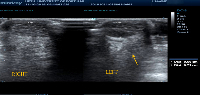
Aim and Objectives: This study was aimed at evaluating the Achilles tendon thickness (ATT) in type 2 diabetic patients comparing the findings to their age and sex-matched non-diabetic counterparts. It also tried to establish a relationship, if any, of ATT with peripheral neuropathy, Body Mass Index (BMI) and duration of disease in the study population.
Methodology: This was an analytical cross-sectional study involving 108 adult diabetic participants and 108 non-diabetic control participants. The ATT in both groups were evaluated by ultrasonography. The ATT of the study group was correlated with their peripheral neuropathy score, BMI and duration of disease. Data was analyzed using Statistical Package for Social Sciences (SPSS) version 20.0 software
Resuts: The mean ATT was higher in diabetic subjects compared to the control subjects. The increased thickness of the AT was significantly more in the presence of PN (p=0.0001). The optimal cut-off point of ATT for identifying the risk of PN in the feet of diabetics was determined to be > 5.75mm with an accuracy of 83.3%
Conclusion: Patients with type 2 diabetes mellitus have significantly thicker AT than their age and sex-matched control subjects and the presence of peripheral neuropathy further worsens the ATT. An ATT of > 5.75mm is the optimal cut-off for identifying the risk of PN in the feet of diabetics.
Downloads
Article Details
Section

This work is licensed under a Creative Commons Attribution-NonCommercial-NoDerivatives 4.0 International License.
The Journal is owned, published and copyrighted by the Nigerian Medical Association, River state Branch. The copyright of papers published are vested in the journal and the publisher. In line with our open access policy and the Creative Commons Attribution License policy authors are allowed to share their work with an acknowledgement of the work's authorship and initial publication in this journal.
This is an open access journal which means that all content is freely available without charge to the user or his/her institution. Users are allowed to read, download, copy, distribute, print, search, or link to the full texts of the articles in this journal without asking prior permission from the publisher or the author.
The use of general descriptive names, trade names, trademarks, and so forth in this publication, even if not specifically identified, does not imply that these names are not protected by the relevant laws and regulations. While the advice and information in this journal are believed to be true and accurate on the date of its going to press, neither the authors, the editors, nor the publisher can accept any legal responsibility for any errors or omissions that may be made. The publisher makes no warranty, express or implied, with respect to the material contained herein.
TNHJ also supports open access archiving of articles published in the journal after three months of publication. Authors are permitted and encouraged to post their work online (e.g, in institutional repositories or on their website) within the stated period, as it can lead to productive exchanges, as well as earlier and greater citation of published work (See The Effect of Open Access). All requests for permission for open access archiving outside this period should be sent to the editor via email to editor@tnhjph.com.
How to Cite
References
Lozano R, Naghavi M, Foreman K, Lim S, Shibuya K, Aboyans V, et al. Global and regional mortality from 235 causes of death for 20 age groups in 1990 and 2010: A systematic analysis for the global burden of disease study 2010. The lancet. 2012;380:2095-2128.
Murray CJ, Vos T, Lozano R, Naghavi M, Flaxman AD, Michaud C, et al. Disability-adjusted life years (dalys) for 291 diseases and injuries in 21 regions, 1990–2010: A systematic analysis for the global burden of disease study 2010. The lancet. 2012;380:2197-2223.
Whiting DR, Guariguata L, Weil C, Shaw J. IDF diabetes atlas: Global estimates of the prevalence of diabetes for 2011 and 2030. Diabetes Res Clin Pract. 2011;94:311-321.
Kengne AP, Amoah AG, Mbanya JC. Cardiovascular complications of diabetes mellitus in sub-Saharan Africa. Circulation. 2005;112:3592-3601.
Akinkugbe OO. Non-communicable diseases in Nigeria: National survey (final report) on hypertension, coronary heart disease, diabetes mellitus, haemoglobinopathy, g6pd deficiency and anaemia national expert committee on non-communication disease. Federal Ministry of Health and Social services. Lagos. 1997.
Guariguata L, Whiting DR, Hambleton I. Global estimates of diabetes prevalence for 2013 and projections for 2035. Diabetes Res Clin Pract 2014;103:137-149.
Chinenye S, Ofoegbu E, Onyemelukwe G, Uloko A, Ogbera A. Clinical practice guidelines for diabetes management in nigeria. Diabetes Association of Nigeria; 2013. Available from: http://gracelanddiabetesfoundation.org/wp-content/uploads/2018/03/Guideline-For-Diabetes-Management-In-Nigeria-2nd-Edition.pdf. [Accessed 3 March 2020].
Nyenwe EA, Odia OJ, Ihekwaba AE, Ojule A, Babatunde S. Type 2 diabetes in adult nigerians: A study of its prevalence and risk factors in Port Harcourt, Nigeria. Diabetes research and clinical practice. 2003;62:177-185.
Hernández-Díaz C, Saavedra MÁ, Navarro-Zarza JE, Canoso JJ, Villasenor-Ovies P, Vargas A, et al. Clinical anatomy of the ankle and foot. Reumatologia clinica. 2012;8:46-52.
Cheung JT, Zhang M, An KN. Effect of achilles tendon loading on plantar fascia tension in the standing foot. Clin Biomech. 2006;21:194-203.
Abate M, Schiavone C, Salini V, Andia I. Occurrence of tendon pathologies in metabolic disorders. Rheumatology. 2013;52:599-608.
Cheing GL, Chau RM, Kwan RL, Choi CH, Zheng YP. Do the biomechanical properties of the ankle–foot complex influence postural control for people with type 2 diabetes? Clin Biomech. 2013;28:88-92.
Frykberg RG, Lavery LA, Pham H, Harvey C, Harkless L, Veves A. Role of neuropathy and high foot pressures in diabetic foot ulceration. Diabetes care. 1998;21:1714-1719.
Rao SR, Saltzman CL, Wilken J, Yak HJ. Increased passive ankle stiffness and reduced dorsiflexion range of motion in individuals with diabetes mellitus. Foot Ankle Int. 2006;27:617-622.
Salsich GB, Mueller MJ, Hastings MK, Sinacore DR, Strube MJ, Johnson JE. Effect of achilles tendon lengthening on ankle muscle performance in people with diabetes mellitus and a neuropathic plantar ulcer. Physical therapy. 2005;85:34-43.
Reddy GK. Glucose‐mediated in vitro glycation modulates biomechanical integrity of the soft tissues but not hard tissues. J Orthop Res. 2003;21:738-743.
Reiber GE, Vileikyte L, Boyko EJ, Del AM, Smith DG, Lavery LA, et al. Causal pathways for incident lower-extremity ulcers in patients with diabetes from two settings. Diabetes Care. 1999;22:157-162.
Papanas N, Courcoutsakis N, Papatheodorou K, Daskalogiannakis G, Maltezos E, Prassopoulos P. Achilles tendon volume in type 2 diabetic patients with or without peripheral neuropathy: Mri study. Exp Clin Endocrinol Diabetes. 2009;117:645-648.
Colen LB, Kim CJ, Grant WP, Yeh JT, Hind B. Achilles tendon lengthening: Friend or foe in the diabetic foot?. Plast Reconstr Surg. 2013;131:37-43.
Zhang P, Lu J, Jing Y, Tang S, Zhu D, Bi Y. Global epidemiology of diabetic foot ulceration: A systematic review and meta-analysis. Ann med. 2017;49:106-116.
Ehusani FE GS, Ohwovoriole AE. A retrospective study of diabetic foot lesion in lagos. Nig J Int Med. 1999:10-12.
Jacobson JA. Ultrasound in sports medicine. Radiol Clin of North Am. 2002;40:363-386.
Rawool NM, Nazarian LN. Ultrasound of the ankle and foot. Semin Ultrasound CT MR. 2000;21:275-284.
Lin J, Fessell DP, Jacobson JA, Weadock WJ, Hayes CW. An illustrated tutorial of musculoskeletal sonography: Part I, introduction and general principles. AJR Am J Roentgenol. 2000;175:637-645.
Erickson S. High-resolution imaging of the musculoskeletal system. Radiology. 1997;205:593-618.
Afolabi BI, Ayoola OO, Idowu BM, Kolawole BA, Omisore AD. Sonographic evaluation of the achilles tendon and plantar fascia of type 2 diabetics in Nigeria. J Med Ultrasound. 2019;27:86-91
Giacomozzi C, D’ambrogi E, Uccioli L, Macellari V. Does the thickening of achilles tendon and plantar fascia contribute to the alteration of diabetic foot loading? Clin biomech. 2005;20:532-539.
Abate M, Schiavone C, Di-Carlo L, Salini V. Achilles tendon and plantar fascia in recently diagnosed type II diabetes: Role of body mass index. Clin Rheumatol. 2012;31:1109-1113.
Akturk M, Ozdemir A, Maral I, Yetkin I, Arslan M. Evaluation of Achilles tendon thickening in type 2 diabetes mellitus. Exp Clin Endocrinol Diabetes. 2007;115:92-96.
Batista F, Nery C, Pinzur M, Monteiro AC, DeSouza EF, Felippe FH, et al. Achilles tendinopathy in diabetes mellitus. Foot Ankle Int. 2008;29(5):498-501.
Evranos B, Idilman I, Ipek A, Polat SB, Cakir B, Ersoy R. Real-time sonoelastography and ultrasound evaluation of the Achilles tendon in patients with diabetes with or without foot ulcers: a cross sectional study. J Diabetes Complications. 2015;29(8):1124-1129.
Gaida JE, Cook JL, Bass S. Adiposity and tendinopathy. Disability and rehabilitation. 2008;30:1555-1562.
Gaida JE, Ashe MC, Bass SL, Cook JL. Is adiposity an under‐recognized risk factor for tendinopathy? A systematic review. Arthritis Rheum. 2009;61:840-849.
Mello RA, Marchiori E, Santos A, Torres N. Morphometric evaluation of achilles tendon by ultrasound. Radiol Bras. 2006;39:161-165.
Tsouli SG, Xydis V, Argyropoulou MI, Tselepis AD, Elisaf M, Kiortsis DN. Regression of achilles tendon thickness after statin treatment in patients with familial hypercholesterolemia: An ultrasonographic study. Atherosclerosis. 2009;205:151-155.
Ursini F, Arturi F, D'Angelo S, Amara L, Nicolosi K, Russo E, et al. High prevalence of achilles tendon enthesopathic changes in patients with type 2 diabetes without peripheral neuropathy. J Am Podiatr Med Assoc. 2017;107:99-105.
Abate M, Salini V, Antinolfi P, Schiavone C. Ultrasound morphology of the achilles in asymptomatic patients with and without diabetes. Foot Ankle Int. 2014;35:44-49.
Teichtahl AJ, Brady SR, Urquhart DM, Wluka AE, Wang Y, Shaw JE, et al. Statins and tendinopathy: A systematic review. Med J Aust. 2016;204:115-121.
Grant WP, Sullivan R, Sonenshine DE, Adam M, Slusser JH, Carson KA, et al. Electron microscopic investigation of the effects of diabetes mellitus on the achilles tendon. The J foot Ankle Surg. 1997;36:272-278.