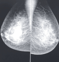
Mammographic Findings Among Women in Port Harcourt: a Multicentre Study
Main Article Content
Abstract
Background: It is the primary imaging modality for breast diseases evaluation, cancer screening and diagnosis. The increasing incidence of breast cancer worldwide has made mammography an important tool; as mammography breast cancer screening had shown to reduce mortality. The aim of the study is to evaluate the indication for mammography referral and the prevalence of breast lesions in our locality with a view to assessing the benefits of mammography practice in our health facilities
Methods: This was a prospective descriptive study of all mammogram performed during the five years of the study. Information on patient’s age, parity, last menstrual period, breastfeeding, occupation, indication for mammogram referral and past mammographic exposure were collected.
Results: The patients’ age ranged from 22 to 78 years with a mean of 48.34 ±1.04 years. Indications for mammography were breast screening for cancer which constituted 97(36.60%) patients, followed by breast lump constituting 74(27.94%) patients and breast pain in 56(21.13%) patients. Evaluation of the mammographic findings showed that 158 (59.62%) patients had normal findings. The BI-RADS category showed high prevalence of category 1 which was found in 155 (58.49%) patients
Conclusion: The study had shown that mammography is an important imaging modality for evaluating breast lesions. Breast screening mammography had become acceptable to women in our society, especially if the financial implication is not burdensome on them. It was also demonstrated that benign breast lesion was commoner than malignant lesion.
Downloads
Article Details
Section

This work is licensed under a Creative Commons Attribution-NonCommercial-NoDerivatives 4.0 International License.
The Journal is owned, published and copyrighted by the Nigerian Medical Association, River state Branch. The copyright of papers published are vested in the journal and the publisher. In line with our open access policy and the Creative Commons Attribution License policy authors are allowed to share their work with an acknowledgement of the work's authorship and initial publication in this journal.
This is an open access journal which means that all content is freely available without charge to the user or his/her institution. Users are allowed to read, download, copy, distribute, print, search, or link to the full texts of the articles in this journal without asking prior permission from the publisher or the author.
The use of general descriptive names, trade names, trademarks, and so forth in this publication, even if not specifically identified, does not imply that these names are not protected by the relevant laws and regulations. While the advice and information in this journal are believed to be true and accurate on the date of its going to press, neither the authors, the editors, nor the publisher can accept any legal responsibility for any errors or omissions that may be made. The publisher makes no warranty, express or implied, with respect to the material contained herein.
TNHJ also supports open access archiving of articles published in the journal after three months of publication. Authors are permitted and encouraged to post their work online (e.g, in institutional repositories or on their website) within the stated period, as it can lead to productive exchanges, as well as earlier and greater citation of published work (See The Effect of Open Access). All requests for permission for open access archiving outside this period should be sent to the editor via email to editor@tnhjph.com.
How to Cite
References
Harold E, Vishy M. Clinical anatomy: Applied anatomy for students and junior doc-tors. Wiley- Blackwell, John Wiley & Sons Ltd publication UK. 12th edition. 2010; 207-211.
Heng D, Gao F, Jong R, Fishell E, Yaffe M, Martin L, et al. Risk factors for breast cancer associated with mammographic features in Singaporean Chinese women. Can-cer Epi Biom Prev. 2004; 13: 1751-8.
Starvos AT, Thickman D, Rapp U, et al. Solid breast nodules: use of sonography to distinguish between benign and malignant lesion. Radiology. 1995; 196: 123-134.
Douglas S. Should we screen women with mammography in Hong Kong? The Hong Kong Practitioner 1994; 16(9): 447-56
Hardy JD, Kukora JS, Harvey IP. Cancer of the breast, epidemiology and aetiology. Hardy’s text book of surgery, JB Lippincott Company Philadelphia, second edition. 1988.
Out AA, Ekanem OO, Khalil ML, Attah EB, Ekpo MD. Characterisation of breast cancer subgroup in an African population. British journal of Surg 1989; 71: 182-4
Okobia MN, Aligbe JU. Pattern of malignant diseases at the University of Benin Teaching Hospital. Trop Doct 2005; 35: 91-2
Tabar L, Vitak B, Chen HH, Prevost TC, Duffy SW. Update of the Swedish two- county trial of breast cancer screening: histological grade-specific and age specific re-sults. Swiss Surg 1999; 5(5): 199-204.
American Cancer Society. Breast cancer facts and figures 2005-2006. Atlanta. Ameri-can Cancer Society; Inc.; 2005.
Akande HJ, Olafimihan BB, Oyinloye OI. A five year audit of mammography in a tertiary hospital, North Central Nigeria. Nigerian Medical Journal 2015; 56(3): 213-17.
Obajimi MO, Adeniji-Sofoluwe AT, Oluwasola AO, Adedokun BO, Soyemi TO, Olopade F et al. Mammographic breast pattern in Nigerian women in Ibadan, Nigeria. Breast Dis. 2011; 33: 9-15
Akinola RA, Akinola OL, Shittu L, Balogun BO, Tayo AO. Appraisal of mammogra-phy in Nigeria women in a new teaching hospital. Scientific Research and Essay 2007; 2(8): 325-9
Ademyomoye AAO, Awosanya GOG, Adesanya AA, Anunobi CC, Osibogun A. Medical audit of diagnostic mammographic examination at the Lagos University Teaching Hospital (LUTH), Nigeria. Niger Postgrad Med J. 2009; 16(1): 25-30.
Eni UE, Ekwedigwe KC, Sunday- Adeoye I, Daniyan ABC, Isikhuemen ME. Audit of mammography requests in Abakaliki, South-East Nigeria. World Journal of Surgical Oncology. 2017; 15: 56. Doi 10.1186/s12957-017-1122-7
Nggad HA, Gali BM, Bakari AA, Tema EH, Tahir MB Apari E, et al. The spectrum of female breast diseases among Nigerian population in Sahel climate zone, J med sci 2011; 2: 115-61
Anele AA, Okoro IO, Oparaocha DC, Igwe PO. Pattern of breast diseases in Owerri, Imo state, Nigeria. Afr J online 2009; 4: 1
Ayoade BA, Tade AO, Salami BA. Clinical features and pattern of presentation of breast diseases in surgical outpatient clinic of a suburban tertiary hospital in South-west Nigeria. Niger J Surg. 2012; 18:13-6
Berta MG, William EB, Rachel BB, Virginia LE, Bonnie CY, Edward AS et al. Use of the American college of radiology BI-RADS to report on the mammographic evaluation of women with signs and symptoms of breast disease. Radiology 2002; 222: 536-542.
Okere P, Aderigbibe A, Iloanusi N, Olusina DB, Itanyi D, Okoye I. An audit of the first three years of mammography and sono-mammography at the University of Nige-ria Teaching Hospital, Enugu, Nigeria. J Coll Med 2012; 17: 2
Ebubedike UR, Umeh EC, Anyanwu SNC, Ukah CO, Ikegwuonu NC. Pattern of mammography findings among symptomatic females referred for diagnostic mammog-raphy at a tertiary center in South-East Nigeria. West African Journal of Radiology 2016; 23(1): 23-7
Ochicha O, Edino ST, Mohammed AZ, Amin SN. Benign breast lesions in Kano, Nig J Surg Res. 2002; 24: 257-62.
Ikpeme A, Akintomide A, Inah G, Oku A. Breast evaluation findings in Calabar, Ni-geria. Macedonian Journal of Medical Sciences 2014; 4(2). Doi 10.3889/oamjms. 2014.117

