
Review of Histological Variants of Uterine Leiomyomas seen in a Teaching Hospital
Article Sidebar
Main Article Content
Abstract
Background: Uterine leiomyoma is the commonest benign smooth muscle tumour of unknown aetiology and occurs mostly in reproductive-age women. The gross appearances are often altered by various degenerative changes and histological types of leiomyomas are of mainly interest as they may mimic malignancy in rare cases. This study described histopathological variants and degenerative changes of uterine leiomyomas seen at LTH, Ogbomoso over a 5- year.
Method: This was a hospital based retrospective study of 192 cases of uterine leiomyoma diagnosed histologically at the Department of Morbid Anatomy LTH, Ogbomoso between January 2012 and December 2016. Cases were retrieved from the histopathology register and demographic characteristics such as age were extracted. Leiomyomas were classified using 2014 edition of World Health Organization Classification of Uterine Smooth Muscle Tumour. Data obtained was analysed using both Microsoft Excel and Statistical Package for Social Sciences 23.0 (SPSS version 23.0).
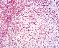
Result: Leiomyoma was commonly seen the fourth decade of life which accounted for about 40.6%. Multiple nodules were found in 136(70.8%) cases. Intramural leiomyoma was the most common site accounting for 176(91.7%) cases. Usual leiomyoma was the commonest histological type accounting for 182(94.8%) cases. Degenerative changes were seen in 124 (64.6%) cases and hyaline change was the commonest secondary change with a frequency of 115 (59.9%).
Conclusion: Majority of women with uterine leiomyomas presented with multiple nodules. Intramural site was the most common location. Hyaline change was the most common degeneration and usual variant was the most common subtype seen in our study.
Downloads
Article Details
Section

This work is licensed under a Creative Commons Attribution-NonCommercial-ShareAlike 4.0 International License.
The Journal is owned, published and copyrighted by the Nigerian Medical Association, River state Branch. The copyright of papers published are vested in the journal and the publisher. In line with our open access policy and the Creative Commons Attribution License policy authors are allowed to share their work with an acknowledgement of the work's authorship and initial publication in this journal.
This is an open access journal which means that all content is freely available without charge to the user or his/her institution. Users are allowed to read, download, copy, distribute, print, search, or link to the full texts of the articles in this journal without asking prior permission from the publisher or the author.
The use of general descriptive names, trade names, trademarks, and so forth in this publication, even if not specifically identified, does not imply that these names are not protected by the relevant laws and regulations. While the advice and information in this journal are believed to be true and accurate on the date of its going to press, neither the authors, the editors, nor the publisher can accept any legal responsibility for any errors or omissions that may be made. The publisher makes no warranty, express or implied, with respect to the material contained herein.
TNHJ also supports open access archiving of articles published in the journal after three months of publication. Authors are permitted and encouraged to post their work online (e.g, in institutional repositories or on their website) within the stated period, as it can lead to productive exchanges, as well as earlier and greater citation of published work (See The Effect of Open Access). All requests for permission for open access archiving outside this period should be sent to the editor via email to editor@tnhjph.com.
How to Cite
References
Okolo S. Incidence, aetiology and epidemiology of uterine fibroids. Best. Pract. Res. Clin. Obstet. Gynaecol. 2008; 22: 571-88.
Hendrickson MR, Tavassoli FA, Kempson RL, Mc Cluggage WG, Haller U, Kubik-Huch RA. Mesenchymal tumours and related lesions. In Tavassoli FA, Deville P(Eds). Pathology and Genetics of Tumours of the Breast and Female Genital Organs. © International agency for research on cancer, Lyon: 2003: 236 – 42.
Maddila Yamuna, D. Hemalatha Devi. Clinical, Sonographical, Surgical, Histopathological study of fibroid. IAIM, 2020;7(2):6-12.
Abraham J, Saldanha P. Morphological variants and secondary changes in uterine leiomyomas – Is it important to recognize them? International Journal of Biomedical Research. 2013;4(12):254–264. Available from: https://dx.doi.org/10.7439/ijbr.v4i12.428.
Sarfraz R, Sarfraz MA, Kamal F, Afsar A. Pattern of benign morphological myometrial lesions in total abdominal hysterectomy specimens. Biomedica. 2010; 26:140-3.
Ukwenya V, Maduemezia N, Afolayan O, Alese O, Thomas W. Prevalence of uterine fibroid in a Southwestern Nigerian population: A sonographic study. J Exp Clin Anat. 2015; 14; 24-9.
Abbas HY, AwadI A, Alharbi E, Alaameri H, Althubaiti S, Ashkar L. Prevalence and incidence of uterine fibroid at King Abdulaziz University Hospital Saudi Arabia. J.CMD 2016; 6: 45-8.
Bizjak T, A TurkanovićAB , But I. Prevalence and risk factors of uterine fibroids in North-East Slovenia. Gynecol Obstet . 2016; 6:1-4.
Lahori M, Malhotra AS, Sakul, Khajuria1 A, Goswami KC. Clinicopathological spectrum of uterine leiomyomas in a state of Northern India: a hospital-based study. Int J Reprod Contracept Obstet Gynecol. 2016; 5:2295-99.
Cramer S F, Patel A. The frequency of uterine leiomyomas. Am. J. Clin. Pathol. 1990; 94: 435-8.
Goyal V, Agrawal R, Mohan N. Analysis of Leiomyoma of Female Genital Tract — Degenerative Changes and Morphological Variants. J Med Sci Health 2021; 7(3):13-18
Gowri M, Mala G, Murthy S, Nayak V. Clinicopathological study of uterine leiomyomas in hysterectomy specimens. J Evol of Med Dent Sci..2013;2(46):9002–9. Available from: https://dx.doi. org/10.14260/jemds/1563.
Manjula K, Kadam SR, Chandrasekhar HR. Variants of leiomyoma: Histomorphological study of tumors of myometrium. JSAFOG 2011; 3:89-92.
Oliva E, Carcangiu ML, Carinelli SG, Ip P, Loening T Longacre TA et al. Mesenchymal tumours. In Kurman RJ, Carcangiu ML, Herrington CS, Young RH and Purcell R(Eds). WHO Classification of Tumours of Female Reproductive Organs. © International agency for research on cancer, Lyon: 2014: 135 – 8.
Mohammed A, Shehu SM, Ahmed SA, Mayun AA, Tiffin IU, Alkali G et al. Uterine leiomyomata: a five-year clinicopathological review in Zaria, Nigeria. Niger J. Surg. Res. 2005; 7: 206-8.
Begum S, Khan S. Audit of leiomyoma uterus at Khyber Teaching Hospital, Peshawar. J Ayub Med Coll.2004;16(2):46–49.
Dayal S, Kumar A, Verma A. Clinicopathologic correlation of leiomyoma with clinical findings and secondary changes in a rural population of North India. American Journal of Clinical Pathology. 2014;141(2):275–279. Available from: https://dx.doi. org/10.1309/ajcpslmz1toc4jcf.ss
Kaur M, Gupta RK, Kaur SJ, Kaur P. Clinicopathological study of leiomyomas in hysterectomy specimens. Int J Reprod Contracept Obstet Gynecol 2018; 7:1509-13.
Flake GP, Andersen J, Dixon D. Etiology and pathogenesis of uterine leiomyomas: A Review. Environ Health Perspect.2003; 111:1037–54.