
Radio-Anatomical Evaluation of the Nasopalatine Canal and its Clinical implication
Article Sidebar
Main Article Content
Abstract
Background: Nasopalatine canal (NPC), also known as incisive canal is in the anterior part of the hard palate posterior to the maxillary incisor and serves as a channel between the oral and nasal cavities. Incisive foramen is the distal opening of the incisive canal on the hard palate. This study evaluated the nasopalatine canal and its morphological variations amongst Nigerians.
Method: This cross-sectional research was done in Radiology department of Rivers State University Teaching Hospital, Nigeria. One hundred computed tomography images of adults (≥18years) were studied. Data were analyzed using SPSS version 22.0. Independent t- test, ANOVA, paired t - test were used for comparison. A p<0.05 was considered statistically significant.
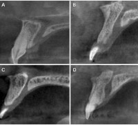
Result: Cylindrical shape of NPC was most prevalent 43(23%), males had more cylindrical shape 38(27.6%), females had more funnel shape 26(24.8%). NPC length for males and females were 16.31+2.904mm and 13.92+2.638mm respectively and is statistically significant. The diameter of the incisive canal was 3.17±1.17mm and 3.24±1.10mm; males and females respectively, not statistically significant. Incisive foramen had no significant correlation as regards age and gender; double foramina was also observed in four female subjects.
Conclusion: Knowledge of this structure and its morphological variations will help surgeons operating in that region in avoiding injury to neurovascular and other related structures. This study also found double foramina in four female subjects.
Downloads
Article Details
Section

This work is licensed under a Creative Commons Attribution-NonCommercial-ShareAlike 4.0 International License.
The Journal is owned, published and copyrighted by the Nigerian Medical Association, River state Branch. The copyright of papers published are vested in the journal and the publisher. In line with our open access policy and the Creative Commons Attribution License policy authors are allowed to share their work with an acknowledgement of the work's authorship and initial publication in this journal.
This is an open access journal which means that all content is freely available without charge to the user or his/her institution. Users are allowed to read, download, copy, distribute, print, search, or link to the full texts of the articles in this journal without asking prior permission from the publisher or the author.
The use of general descriptive names, trade names, trademarks, and so forth in this publication, even if not specifically identified, does not imply that these names are not protected by the relevant laws and regulations. While the advice and information in this journal are believed to be true and accurate on the date of its going to press, neither the authors, the editors, nor the publisher can accept any legal responsibility for any errors or omissions that may be made. The publisher makes no warranty, express or implied, with respect to the material contained herein.
TNHJ also supports open access archiving of articles published in the journal after three months of publication. Authors are permitted and encouraged to post their work online (e.g, in institutional repositories or on their website) within the stated period, as it can lead to productive exchanges, as well as earlier and greater citation of published work (See The Effect of Open Access). All requests for permission for open access archiving outside this period should be sent to the editor via email to editor@tnhjph.com.
How to Cite
References
Venkatesh E, Elluru SV. Cone beam computed tomography: basics and applications in dentistry. J Istanb Fac Dent. 2017;;51(3):102-121, doi: 10.17096/jiuf
Salemi F, Moghadam FA, Shakibai Z, Farhadian M. Three-dimensional assessment of the nasopalatine canal and the surrounding bone using cone-beam computed tomography. J Periodontol Implant Dent. 2016;8(1):1-7.
Bornstein MM, Balsiger R, Sendi P, von Arx T. Morphology of the nasopalatine canal and dental implant surgery: A radiographic analysis of 100 consecutive patients using limited cone-beam computed tomography. Clin Oral Implants Res; 2011; 22:295-301.
Asaumi R, Kawai T, Sato I, Yoshida S, Yosue T. Three-dimensional observations of the incisive canal and the surrounding bone using cone-beam computed tomography. Oral Radiol 2010; 26:20-8.
Chatriyanuyoke P, Lu CI, Suzuki Y, Lozada JL, Rungcharassaeng K, Kan JY, et al. Nasopalatine canal position relative to the maxillary central incisors: a cone beam computed tomography assessment. J Oral Implantol. 2012;38(6):713-717.
Safi Y, Moshfeghi M, Rahimian S, Kheirkhahi M, Manouchehri ME. Assessment of nasopalatine canal anatomic variations using cone beam computed tomography in a group of Iranian population. Iran J Radiol. 2017;14(1)e37028
Etoz M, Sisman Y. Evaluation of the nasopalatine canal and variations with cone-beam computed tomography. Surg Radiol Anat. 2014;36(8):805-812
Nemtoi, A., Sirghe, A. E., Nemtoi, A, Dobrovat, B., Popescu, R. and Haba, D. CBCT analysis of the morphology and anatomical variants of the nasopalatine canal in northeastern population of Romania. Romanian journal of oral rehabilitation. 2019;11(4), 213-227.
Milanovic, P & Vasiljevic. Gender differences in the morphological characteristics of the nasopalatine canal and anterior maxillary bone-CBCT study. Experimental and applied biomedical research, 1-10, doi: https//doi.org/10.2478/sjecr-2021-0029
Hakbilen S, Magat G. Evaluation of anatomical and morphological characteristics of the nasopalatine canal in a Turkish population by cone beam computed tomography. Folia Morphol (Warsz). 2018; 77(3): 527-535.
Kajan ZD, Kia J, Motevasseli S, Rezaian SR. Evaluation of the nasopalatine canal with cone-beam computed tomography in an Iranian population. Dent Res J (Isfahan). 2015;12(1):14-19. doi: 10.4103/1735-3327.150289.
Thakur AR, Burde K, Guttal K, Naikmasur VG. Anatomy and morphology of the nasopalatine canal using cone-beam computed tomography. Imaging Sci Dent. 2013;43(4):273-281.
Sekerci AE, Buyuk SK, Cantekin K. Cone-beam computed tomographic analysis of the morphological characterization of the nasopalatine canal in a pediatric population. Surg Radiol Anat. 2014;36(9):925-932.
Fernández-Alonso A, Suárez-Quintanilla JA, Muinelo-Lorenzo J, Varela-Mallou J, Smyth Chamosa E, Suárez-Cunqueiro MM. Critical anatomic region of nasopalatine canal based on tridimensional analysis: cone beam computed tomography. Sci Rep. 2015; 5:12568.
Panda M, Shankar T, Raut A, Dev S, Kar AK, Hota S. Cone beam computerized tomography evaluation of incisive canal and anterior maxillary bone thickness for placement of immediate implants. J Indian Prosthodont Soc.2018; 18(4): 356-.363.
Bornstein MM, Balsiger R, Sendi P, von Arx T. Morphology of the nasopalatine canal and dental implant surgery: A radiographic analysis of 100 consecutive patients using limited cone-beam computed tomography. Clin Oral Implants Res; 2011; 22:295-301
Liang X, Jacobs R, Martens W, Hu Y, Adriaensens P, Quirynen M, et al. Macro and micro‐anatomical, histological and computed tomography scan characterization of the nasopalatine canal. J Clin Periodontol. 2009; 36(7):598-603.
Mardinger O, Namani-Sadan N, Chaushu G, Schwartz-Arad D. Morphologic changes of the nasopalatine canal related to dental implantation: a radiologic study in different degrees of absorbed maxillae. J Periodontol. 2008;79(9):1659-1662
Artzi Z, Nemcovsky CE, Bitlitum I, Segal P. Displacement of the incisive foramen in conjunction with implant placement in the anterior maxilla without jeopardizing vitality of nasopalatine nerve and vessels: A novel surgical approach. Clin Oral Implants Res; 2000; 11:505-10.
Linjawi, A., et al. (2021). "Morphological evaluation of the incisive canal with reference to gender and age: A cone-beam computed tomography study." Nigerian Journal of Clinical Practice. 2021; 24(11): 1596.
Mohammed, D. A. Anatomical variation in the dimension of nasopalatine canal on cone beam computed tomography CBCT images among Sulaimani population. Acta Medica Internation. 2016; 3(2): 82-87, Doi:10.5530/ami.2016.2.18
Sicher H. (1962). Anatomy and oral pathology. Oral Surg Oral Med Oral Pathol. 15:1264–9.
Jacobs R, Lambrichts I, Liang X, Martens W, Mraiwa N, Adriaensens P, et al. Neurovascularization of the anterior jaw bones revisited using high-resolution magnetic resonance imaging. Oral Surg Oral Med Oral Pathol Oral Radiol Endod.2007;103(5):683–93.
Song WC, Jo DI, Lee JY, Kim JN, Hur MS, Hu KS, et al. Microanatomy of the incisive canal using three-dimensional reconstruction of microCT images: an ex vivo study. Oral Surg Oral Med Oral Pathol Oral Radiol Endod. 2009;108(4):583–90. doi: 10.1016/j.tripleo.2009.06.036
Soumya P, Koppolu P, Pathakota KR, Chappidi V. Maxillary incisive canal characteristics: a radiographic study using cone beam computerized tomography. Radiology research and practice. 2019;2019(1):6151253.