MRI-Based Morphometric Study of the Corpus Callosum in Nigerian Adults
Main Article Content
Abstract
Background: Alterations in the dimensions of the corpus callosum have been linked to conditions like Alzheimer’s, schizophrenia and bipolar disorder. This study was designed to investigate the normal cut-off measurements of the corpus callosum.
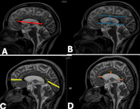
Methods: The cross-sectional study retrospectively assessed the corpus callosum on apparently normal brain Magnetic Resonance scans of 199 adults (91 males, 108 females) aged 20-80 years. The digital radiological storage of a Nigerian Hospital in Delta State was accessed for data collection after institutional authorization was granted. Statistical Package for Social Sciences analyzed the gathered data using the independent t-test for sex comparison and analysis of variance for age-related differences. Inter-variable association was probed using the Pearson’s correlation test. Significance in the inferential statistics was deemed at P<0.05.
Results: Sexual dimorphism was observed in the distance of the callosum’s genu and splenium from the frontal and occipital poles of the brain respectively (p= 0.010 and 0.007). The corpus callosum’s length, height, index, and thickness of its genu and splenium exhibited significant disparities across age-groups. The length and height had a positive association with age while the thickness of the splenium had a negative correlation with age (p<0.05).
Conclusion: This study provides normative range morphometric values for the normal corpus callosum in adults that will aid in accurate diagnosis and follow-up of neurodegenerative and psychiatric conditions besides planning for corpus callosotomy in epileptic patients.
Downloads
Article Details
Section

This work is licensed under a Creative Commons Attribution-NonCommercial-ShareAlike 4.0 International License.
The Journal is owned, published and copyrighted by the Nigerian Medical Association, River state Branch. The copyright of papers published are vested in the journal and the publisher. In line with our open access policy and the Creative Commons Attribution License policy authors are allowed to share their work with an acknowledgement of the work's authorship and initial publication in this journal.
This is an open access journal which means that all content is freely available without charge to the user or his/her institution. Users are allowed to read, download, copy, distribute, print, search, or link to the full texts of the articles in this journal without asking prior permission from the publisher or the author.
The use of general descriptive names, trade names, trademarks, and so forth in this publication, even if not specifically identified, does not imply that these names are not protected by the relevant laws and regulations. While the advice and information in this journal are believed to be true and accurate on the date of its going to press, neither the authors, the editors, nor the publisher can accept any legal responsibility for any errors or omissions that may be made. The publisher makes no warranty, express or implied, with respect to the material contained herein.
TNHJ also supports open access archiving of articles published in the journal after three months of publication. Authors are permitted and encouraged to post their work online (e.g, in institutional repositories or on their website) within the stated period, as it can lead to productive exchanges, as well as earlier and greater citation of published work (See The Effect of Open Access). All requests for permission for open access archiving outside this period should be sent to the editor via email to editor@tnhjph.com.
How to Cite
References
1. Pasricha N, Sthapak E, Thapar A, Bhatnagar R. Morphometric Analysis of Age and Gender-related Variations of Corpus Callosum by using Magnetic Resonance Imaging: A Cross-sectional Study. JCDR. 2023;17(6):15-20.
2. Ajare EC, Campbell FC, Mgbe EK, Efekemo AO, Onuh AC, Nnamani AO, Okwunodulu O, Ohaegbulam SC. MRI-based morphometric analysis of corpus callosum dimensions of adults in Southeast Nigeria. LJM. 2023;18(1):2188649.
3. Arda KN, Akay S. The relationship between corpus callosum morphometric measurements and age/gender characteristics: a comprehensive MR imaging study. JCIS. 2019; 9:33.
4. Choudhury PR, Choudhury P, Baruah P. Morphometric Assessment of Human Corpus Callosum on Cadaveric Brain Specimens. J Evol Med Dent Sci. 2020;9(7):388-393.
5. Poleneni SR, Jakka LD, Chandrupatla M, Vinodini L, Ariyanachi K. Morphometry of Corpus Callosum in South Indian Population. Ann Neurosci. 2021;28(3-4):150-155.
6. Al-Hashim AH, Blaser S, Raybaud C, MacGregor D. Corpus callosum abnormalities: neuroradiological and clinical correlations. DMCN. 2016;58(5):475–484.
7. Tanaka-Arakawa MM, Matsui M, Tanaka C, Uematsu A, Uda S, Miura K, Sakai T, Noguchi K. Developmental changes in the corpus callosum from infancy to early adulthood: a structural magnetic resonance imaging study. PLoS One. 2015;10(3):e0118760.
8. Al-Hadidi MT, Kalbouneh HM, Ramzy A, Al Sharei A, Badran DH, Shatarat A, Tarawneh ES, Mahafza WS, Al-Hadidi FA, Hadidy AM. Gender and age-related differences in the morphometry of corpus callosum: MRI study. Eur J Anat. 2021;25(1):15-24
9. Sehgal G, Kumar A, Kumar G, Aggarwal N. Gender-related differences in the morphometry of the corpus callosum: MRI study. Asian J Med Sci. 2023;14(9):1-6.
10. Habibi HA, Gül OB, Caliskan E, Öztürk M. Morphometric Analysis of Corpus Callosum with ID Magnetic Resonance Imaging in Children; Correlation with Age and Gender. J Behcet Uz Child Hosp. 2021;11(3):277-285.
11. Tuncer I. Morphometric Evaluation of the Human Corpus Callosum using Magnetic Resonance Imaging: Sex Difference and Relationship to Age and Intracranial Size. JASI. 2023;72(2):114-121.
12. Jidong DE, Husain MI, Ike TJ, Husain N, Taru MY, Nnaemeka NC, Francis C, Jack DB, Mwankon SB, Xue S, Pwajok JY. Bipolar disorders in Nigeria: a mixed-methods study of patients, family caregivers, clinicians, and the community members perspectives. Int J Bipolar Disord. 2023;11(1):1-9.
13. Patra A, Singla R, Chaudhary P, Malhotra V. Morphometric analysis of the corpus callosum using cadaveric brain: an anatomical study. Asian J Neurosurg. 2020;15(2):322-327.
14. Ominde BS, Ogbolu EC, Ikubor JE, Omoro OF, Igbigbi PS. Morphometric analysis of the frontal horns of the lateral ventricles using normal computed tomographic images. Eur J Anat. 2024;28(5):615-621.
15. Khasawneh RR, Abu-El-Rub E, Alzu’bi A, Abdelhady GT, Al-Soudi HS. Corpus callosum anatomical changes in Alzheimer patients and the effect of acetylcholinesterase inhibitors on corpus callosum morphometry. PLoS One. 2022;17(7):e0269082.
16. Mastery Farahani R, Aliaghaei A, Abdolmaleki A, Abbaszadeh HA, Shaerzadeh F, Norozian M, Moayeri A. Sexual Dimorphism and Age-Related Variations of Corpus Callosum Using Magnetic Resonance Imaging. Anat Sci J. 2017;14(1):19-26.
17. Smyth MD, Vellimana AK, Asano E, Sood S. Corpus callosotomy—Open and endoscopic surgical techniques. Epilepsia. 2017; 58:73-79.
18. Mohammed A, Ibrahim A, Mohammed ME, Ayad CE. A Quantitative MRI Study of the Normative Corpus Callosum in Sudanese. IOSR-JDMS. 2017;16(7):77-86.
19. Abdolmaleki A, Mastery FR, Ghoreishi SK, Shaerzadeh F, Aliaghaei A, Mirjavadi SH, Abbaszadeh H. Magnetic resonance imaging-based morphometric assessment of sexual dimorphism of corpus callosum. Anat Sci J. 2016;13(2):117-124
20. Tepper R, Leibovitz Z, Garel C, Sukenik-Halevy R. A New Method for Evaluating Short Fetal Corpus Callosum. Prenat Diagn. 2019;39(13):1283-90.

